
Facilities
By measuring the small changes in electrical activity of the brain from the scalp, electroencephalography (or EEG) has provided insights into the temporal sequence of perceptual, cognitive, and motor functions for decades. From within an electrically-shielded room, researchers in the EEG Lab can measure brain activity from up to 64 electrodes on the head, while simultaneously recording behavioral responses and eye tracking.
Technical details
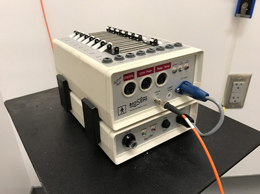
The EEG Lab is located in room 405G. It houses an ActiveTwo Biosemi EEG System, a high impedance system capable of collecting in a 64-Channel configuration. The EEG Lab is one room with a display computer and collection computer. Various devices are available to the subject for behavioral responses. We have a Labhackers button box for collecting millisecond accurate responses and a MilliKey DeLux Light Sensor to detect changes in experiment display brightness and generate a keyboard or serial event when the light level crosses a configurable digital threshold. This information can be used to calculate the difference between reported and actual stimulus onset times.
Tracking eye movements allows us to follow along the path of attention deployed by the observer, giving us insight into what they found interesting or how they perceived whatever is being viewed.
Technical details
The laboratory houses two Tobii Pro Spectrum systems (in rooms 405G and 405F).
- This system is a screen-based eye tracker capturing gaze data at speeds up to 600 Hz. This high-performance research system provides superior data quality and is designed for extensive research into behavior and eye movements – from fixation-based studies to micro-saccades. It combines video-based pupil- and corneal reflection eye tracking with dark and bright pupil illumination modes.
- Two cameras capture stereo images of both eyes for robust accurate measurement of eye gaze and eye position in 3D space, as well as pupil diameter. Sampling frequencies range between 60-600 Hz with precision and accuracy between 0.01-0.06°(RMS), and 0.3°, respectively. The system provides binocular eye tracking with total system latency of less than 3 frames and a blink recovery time of 1 frame. The dual-camera, relative to one camera, system gives a more accurate data calculation and the best level of precision and robustness for head movement.
- The hardware is combined with Tobii Pro Lab software which provides a visual user interface and software features that efficiently guide and support the researcher through all phases of an eye tracking experiment – from test design and recording to analysis. The Tobii Pro Lab facilitates the combination of eye tracking with biometric data streams, like EEG, GSR, and ECG. For co-registration with other data sources, the software allows for a TTL marker at every stimulus onset to ensure precise data synchronization.
- Lab-based studies, including experiments implemented with PsychoPy
- Naturalistic eye-tracking in classrooms, conversations, and real-world navigation
- Multimodal experiments combining eye tracking with EEG or behavioral measures
Measuring participants’ decisions, reaction times and accuracy are integral aspects of human neuroscience research. One room is available to researchers interested in conducting behavioral experiments.
While most human neuroscience methods allow researchers to correlate brain activity with behavior, with transcranial magnetic stimulation (or TMS) researchers can directly manipulate neural activity in a safe manner. TMS can induce virtual “lesions” for brief windows of time to produce short-lasting behavioral or perceptual deficits.
Technical details
We employ a state-of-the-art system for transcranial magnetic stimulation (TMS). The system includes (1) The Magstim Rapid² repetitive stimulator with high frequency capabilities and can also be used for single-pulse protocols (stimulation frequency up to 36Hz (11Hz at max power) (2) AirFilm Butterfly Coil capable of delivering repetitive pulses at high power levels, the D70 AirFilm Coil features an air-cooling system, and (3) a MEP (Motor Evoked Potentials) Monitor. The AFC’s fan permits minimal down time between the running of high intensity protocols, such as high-frequency repetitive pulse protocols.
A Brainsight 2 Neuronavigation System (Rogue Research, Montreal, Canada) is used to position the TMS coil to target specific brain areas. This technology uses procedures developed for pre-surgical planning. Markers on the coil are imaged with cameras and positioned based on the subject’s own anatomy. An LCD monitor is available for stimulus presentation and software is available for precise time locking of the task paradigm to stimulus presentation.
By measuring the changes in cerebral blood flow and oxygenation in the brain, functional near-infrared spectroscopy (fNIRS) has become a valuable tool for studying neural activity associated with perceptual, cognitive, and motor functions. Using near-infrared light, fNIRS allows researchers to non-invasively monitor hemodynamic responses in the brain, offering insight into localized brain activity. The fNIRS system in our lab can simultaneously measure oxygenated and deoxygenated hemoglobin concentrations from multiple optodes positioned on the scalp. This enables the exploration of neural dynamics in both task-related and naturalistic environments, complementing other behavioral and physiological measures such as eye tracking and motion capture.
Technical details
The lab is located in McGuinn 405D
System Features: The Brite24 system measures, stores, and wirelessly transmits up to 27 channels of fNIRS signals. Channels can be configured for large regions of the scalp or subdivided into smaller regions, offering flexibility:
Configurations:
Single large region: 27 channels
Combinations of smaller regions: 2×12 channels, 2×10 + 2 short channels, or 4×4 + 2 short channels.
Optode Setup: The system uses 10 transmitters and 8 receivers that fit into a full-head cap with 64+ optode positions, allowing for extensive placement customization.
Head Cap Options: The lab is equipped with three cap sizes (small, medium, and large) to accommodate different head sizes.
Synchronization Capabilities:
The PortaSync wireless device integrates seamlessly with the Brite24 system, enabling synchronization with other instruments. It can generate dual signal levels, corresponding to different event types, which are recorded alongside fNIRS data.Software:
OxySoft: Used for data collection, storage, and analysis, OxySoft provides real-time and parallel recording of multiple devices. It offers customizable configurations for individual research needs and supports online visualization and analysis.
EMSE Suite: This modular system includes tools such as the Data Editor Module, which supports filtering, transformation, and detailed time-series analysis.
This versatile setup allows researchers to design experiments with exceptional precision, synchronize multimodal datasets, and analyze neural activity across a variety of contexts and conditions.